Tag Archives: neuromas

Temporal changes in mechanical nociceptive thresholds in juvenile pigs subjected to surgical tail amputation: a model of injury induced by tail biting

Temporal changes in mechanical nociceptive thresholds in juvenile pigs subjected to surgical tail amputation: a model of injury induced by tail biting. By Di Giminiani, P., E. Malcolm, M. Leach, M. Herskin, D. Sandercock, S. Edwards, 2016. Royal Dublin Society: Abstracts book of the 24th International Pig Veterinary Society (IPVS) Congress, Dublin, Republic of Ireland 7-10th June 2016. p. 649.

Abstract

Introduction: Tail biting is a global welfare problem in the pig industry leading to significant tail injury and potential carcass rejection. The temporal effects of such injuries and subsequent healing are presently unknown, although limb amputation in humans can lead to abnormal neural activity and decreased nociceptive thresholds. In order to evaluate potential sensitisation following tail damage, we created a model by surgical amputation of tails, and assessed mechanical nociceptive thresholds.

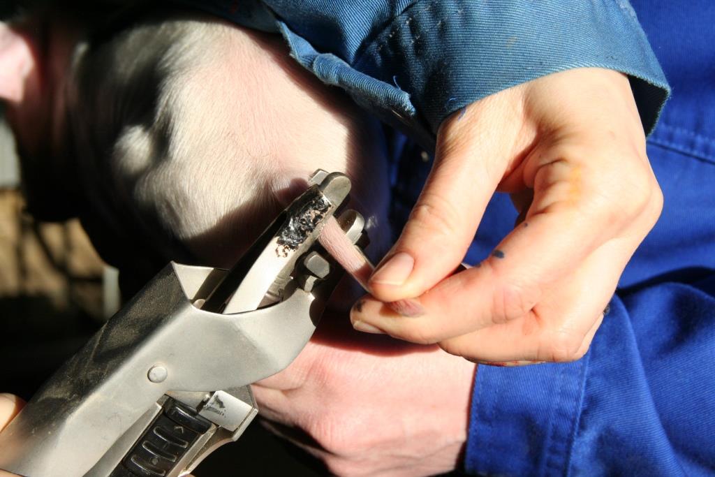
Materials and Methods: Surgical tail resection was performed to assess the influence of age, extent of tail amputated and time since amputation on thresholds of mechanical nociception. To evaluate the effect of age at the time of injury, female pigs underwent surgery at 9 weeks (±3 days ‘weaner’) (n=19) or 17 weeks (±3 days ‘finisher’) (n=43). The effect of time after amputation was evaluated on 24 pigs at 8 weeks, and 38 pigs at 16 weeks after surgery. The effect of the extent of tail amputated was assessed by assigning the pigs to 3 treatments (‘Intact’: sham-amputation; ‘short tail’: 2/3 of tail removed; ‘long tail’: 1/3 of tail removed). A Pressure Application Measurement device was used to record mechanical nociceptive thresholds (tail flick or tail clamp withdrawal responses). Within a single session, three stimuli were applied to a skin area proximal to the site of amputation, 3 days pre-surgery, 1 week and either 8 or 16 weeks post-amputation.

Results: Across the two amputation ages, results indicated that tail amputation induced a significant reduction (P<0.05) in mechanical nociceptive thresholds in short and long tails one week after surgery. The same treatment effect was observed at 16 weeks after amputation performed at 9 weeks of age (P<0.05). For surgeries performed at 17 weeks of age, thresholds tended to be lower in short compared to intact tails (P=0.081) and significantly lower (P<0.05) in long tail pigs 8 weeks after amputation. No significant difference was observed at 16 weeks following surgeries performed at 17 weeks of age.

Conclusion: These results show that surgical amputation of pig tails leads to decreased cutaneous mechanical nociceptive thresholds in the skin area proximal to the site of injury. Results indicated that severe tail injury occurring in the weaner period may be associated with sensitisation up to 16 weeks following the injury. In contrast, injuries occurring in the finishing period appeared to be associated with shorter lasting mechanical sensitisation, resolving within 16 weeks.

Poster Di Giminiani IPVS


Application of a handheld Pressure Application Measurement device for the characterisation of mechanical nociceptive thresholds in intact pig tails

Application of a handheld Pressure Application Measurement device for the characterisation of mechanical nociceptive thresholds in intact pig tails. By Pierpaolo Di Giminiani, Dale A. Sandercock, Emma M. Malcolm, Matthew C. Leach, Mette S. Herskin and Sandra A. Edwards. 2016. Physiology & Behavior 165: 119–126.

Highlights

• Mechanical nociceptive thresholds were quantified for the first time in pig tails.
• The PAM device allowed determining anatomical and age-specific thresholds in pigs.
• A platform for the assessment of painful conditions in pigs is proposed.

Abstract

The assessment of nociceptive thresholds is employed in animals and humans to evaluate changes in sensitivity potentially arising from tissue damage. Its application on the intact pig tail might represent a suitable method to assess changes in nociceptive thresholds arising from tail injury, such as tail docking or tail biting. The Pressure Application Measurement (PAM) device is used here for the first time on the tail of pigs to determine the reliability of the methods and to provide novel data on mechanical nociceptive thresholds (MNT) associated with four different age groups (9, 17, 24 and 32 weeks) and with proximity of the target region to the body of the animal. We recorded an overall acceptable level of intra-individual reliability, with mean values of CV ranging between 30.1 and 32.6%. Across all age groups, the first single measurement of MNT recorded at region 1 (proximal) was significantly higher (P b 0.05) than the following two. This was not observed at tail regions 2 and 3 (more distal). Age had a significant effect (P b 0.05) on the mean thresholds of nociception with increasing age corresponding to higher thresholds. Furthermore, a significant effect of proximity of tail region to the body was observed (P b 0.05), with MNT being higher in the proximal tail region in pigs of 9, 17 and 24 weeks of age.
There was also a significant positive correlation (P b 0.05) between mechanical nociceptive thresholds and age/body size of the animals.
To the best of our knowledge, no other investigation of tail nociceptive thresholds has been performed with the PAM device or alternative methods to obtain mechanical nociceptive thresholds in intact tails of pigs of different age/body size. The reliability of the data obtained with the PAM device support its use in the measurement of mechanical nociceptive threshold in pig tails. This methodological approach is possibly suitable for assessing changes in tail stump MNTs after tail injury caused by tail docking and biting.

Histopathological Characterization of Tail Injury and Traumatic Neuroma Development after Tail Docking in Piglets

Histopathological Characterization of Tail Injury and Traumatic Neuroma Development after Tail Docking in Piglets. By: D.A. Sandercock, S.H. Smith, P. Di Giminiani, S.A. Edwards, 2016. Journal of Comparative Pathology 155: 40-49.

Abstract

Tail docking of neonatal pigs is widely used as a measure to reduce the incidence of tail biting, a complex management problem in the pig industry. Concerns exist over the long-term consequences of tail docking for possible tail stump pain sensitivity due to the development of traumatic neuromas in injured peripheral nerves. Tail stumps were obtained post mortem from four female pigs at each of 1, 4, 8 and 16 weeks following tail amputation (approximately two-thirds removed) by a gas-heated docking iron on post natal day 3. Tissues were processed routinely for histopathological examination. Non-neural inflammatory and reparative epidermal and dermal changes associated with tissue thickening and healing were observed 1 to 4 months after docking. Mild neutrophilic inflammation was present in some cases, although this and other degenerative and non-neural reparative changes are not likely to have caused pain. Traumatic neuroma and neuromatous tissue development was not observed 1 week after tail docking, but was evident 1 month after tail docking. Over time there was marked nerve sheath and axonal proliferation leading to the formation of neuromata, which were either localized and circumscribed or comprised of multiple axons dispersed within granulation tissue. Four months after tail resection, neuroma formation was still incomplete, with possible implications for sensitivity of the tail stump.

Corrigendum to “Histopathological Characterization of Tail Injury and Traumatic Neuroma Development after Tail Docking in Piglets” J Comp Pathol 155 (1) (2016) 40-49.

The authors wish to clarify terminology used in their paper entitled ‘Histopathological characterization of tail injury and traumatic neuroma development after tail docking in piglets’ and thank the Editor for the opportunity to do so. In the absence of a specific immunohistochemical label for detection of axons, the words ‘axon/axonal’ were inaccurately used and should be replaced by ‘Schwann cell’. Without more specific proof, it
certainly does not confirm, or necessarily infer, conduction. Secondly, ‘S100 neurofilament’ was inadvertently used instead of simply ‘S100’. The authors apologise for this error, which was wholly editorial on their part. Finally, in our opinion, the literature definitions of traumatic neuromas are such that there is likely to be some disagreement as to their required component features, particularly at different stages of lesion development, in different species and in different age groups of animals. In our paper, descriptions of traumatic neuroma presence and development were also based on haematoxylin and eosin staining and not solely confined to S100 immunolabelling. To this end, features such as variably-sized microfascicles, disorderly (often circumferential) neural proliferation and nerve fibres turning back on themselves are consistent with previous reports on traumatic neuromas in a number of species, including pigs.
While the aforementioned errors are regretted, this work was intended as a descriptive morphological characterization of a wide range of histopathological changes over known time points post docking. We have had some criticism that, due to our use of the word axonal, we have implied or claimed innervation, and thus pain sensation,
during the weeks after docking. This was not our intention – rather, our opinion is neutral in terms of whether or not traumatic neuromas are painful. The last sentence of the paper acknowledged that this work could not determine that. This study was considered descriptive and foundational, to serve as a platform for further investigation.
Its take-home message, irrespective of the error in terminology, is that neural proliferation consistent with traumatic neuroma development appears to be still ongoing at 16 weeks after tail docking.

Effects of tail docking and docking length on neuroanatomical changes in healed tail tips of pigs

Effects of tail docking and docking length on neuroanatomical changes in healed tail tips of pigs
By M. S. Herskin, K. Thodberg and H. E. Jensen. 2015. Animal 9: 677-681.

Abstract

In pig production, piglets are tail docked at birth in order to prevent tail biting later in life. In order to examine the effects of tail docking and docking length on the formation of neuromas, we used 65 pigs and the following four treatments: intact tails (n=18); leaving 75% (n=17); leaving 50% (n=19); or leaving 25% (n=11) of the tail length on the pigs. The piglets were docked between day 2 and 4 after birth using a gas-heated apparatus, and were kept under conventional conditions until slaughter at 22 weeks of age, where tails were removed and examined macroscopically and histologically. The tail lengths and diameters differed at slaughter (lengths: 30.6±0.6; 24.9±0.4; 19.8±0.6; 8.7±0.6 cm; P<0.001; tail diameter: 0.5±0.03; 0.8±0.02; 1.0±0.03; 1.4±0.04 cm; P<0.001, respectively). Docking resulted in a higher proportion of tails with neuromas (64 v. 0%; P<0.001), number of neuromas per tail (1.0±0.2 v. 0; P<0.001) and size of neuromas (1023±592 v. 0 μm; P<0.001). The results show that tail docking piglets using hot-iron cautery causes formation of neuromas in the outermost part of the tail tip. The presence of neuromas might lead to altered nociceptive thresholds, which need to be confirmed in future studies.

Histological and neurophysiological pain assessment in young pigs

Original title: Approche histologique et neurophysiologie de la douleur liée à la coupe de queue chez les porcelets

Presentation of Dr. Dale Sandercock (SRUC) at a seminar on histological and neurophysiological approaches to pain assessment in young pigs. INRA-PEGASE, St-Gilles, France, December 14th 2015

Abstract

Concerns exist over the long term consequences of tail docking on possible tail stump pain sensitivity due to the development of traumatic neuromas in injured peripheral nerves. Traumatic neuroma formation may cause detrimental sensory changes in the tail due to altered peripheral and spinal neuronal excitability leading to abnormal sensation or pain. We have investigated tail injury and traumatic neuroma development by histopathological assessment after tail docking and measured the expression of key neuropeptides associated with peripheral nerve regeneration, inflammation and chronic pain. In complimentary studies on tail docking and tail biting, we have developed behavioural assessment approaches to measure mechanical nociceptive thresholds (MNT) in the pig tail in purpose built test set-up using a Pressure Application Measurement (PAM) device. Using these approaches we have determined baseline MNT in intact tails along different regions of the tail and also measured changes in MNT over time in pig with resected tails (simulation of the effect of tail biting). An overview of other Scotland’s Rural College (SRUC) pig health and welfare research projects is also presented.

Presentation Pic INRA seminar on pain (D Sandercock, 2015))

Neuroanatomical changes in pig tails following tail docking

Herskin et al. (2015) studied the formation of neuroma’s in pigs after tail docking.

Abstract

In pig production, piglets are tail docked at birth in order to prevent tail biting later in life. In order to examine the effects of tail docking and docking length on the formation of neuromas, we used 65 pigs and the following four treatments: intact tails (n=18); leaving 75% (n=17); leaving 50% (n=19); or leaving 25% (n=11) of the tail length on the pigs. The piglets were docked between day 2 and 4 after birth using a gas-heated apparatus, and were kept under conventional conditions until slaughter at 22 weeks of age, where tails were removed and examined macroscopically and histologically. The tail lengths and diameters differed at slaughter (lengths: 30.6±0.6; 24.9±0.4; 19.8±0.6; 8.7±0.6 cm; P<0.001; tail diameter: 0.5±0.03; 0.8±0.02; 1.0±0.03; 1.4±0.04 cm; P<0.001, respectively). Docking resulted in a higher proportion of tails with neuromas (64 v. 0%; P<0.001), number of neuromas per tail (1.0±0.2 v. 0; P<0.001) and size of neuromas (1023±592 v. 0 μm; P<0.001). The results show that tail docking piglets using hot-iron cautery causes formation of neuromas in the outermost part of the tail tip. The presence of neuromas might lead to altered nociceptive thresholds, which need to be confirmed in future studies.

Sources

Herskin, M.S., Thodberg, K., Jensen, H.E. 2015. Effects of tail docking and docking length on neuroanatomical changes in healed tail tips of pigs. Animal 9: 677-681.
Tail docking causes neuroanatomical changes to pig tails, PigProgress, 25-3-2015

Tail docking using hot iron cautery

Training course animal welfare in pig production

Dr. Dale Sandercock recently took part as a tutor along with Dr. Mette Herskin and Dr. Lene Juul Pedersen in an EU – Better Training for Safer Food (BTSF) training course on Animal Welfare in Pig Production in Herning, Denmark (12-15 May, 2014). The course was aimed at Official Veterinarians belonging to the NCAs (National Competent Authorities) of EU countries involved in official controls on animal welfare concerning the farming of pigs and the development of best practices to improve the application of EU standards. Dale presented a lecture on mutilation procedures: Welfare implications and new strategies. As part of the presentation he reported on the recent setting up of the FareWellDock project and it’s aims in relation to the issues of tail docking and tail biting. Dale was approached by several of the participants for more information about the project and he directed them to this website for further information. The course was attended by 53 participants (mostly from national veterinary institutes) from 28 EU, and candidate countries.

Traning course on mutilations by Dr. Dale Sandercock
Training course on mutilations by Dr. Dale Sandercock